Affiniti 70
Ultrasound system


Affiniti 70
Ultrasound system
Affiniti 70 Elevate is the most advanced system in the Affiniti family, delivering stunning image quality and a suite of premium clinical features. Offering new levels of diagnostic confidence and reproducibility, it is designed for fast-paced environments with enhanced workflow and robust performance—helping you deliver the best possible care every day.
Clinical image gallery
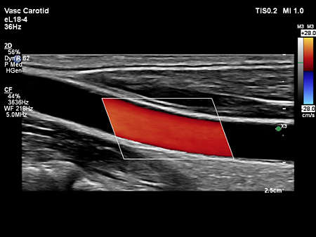
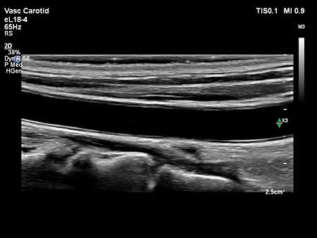
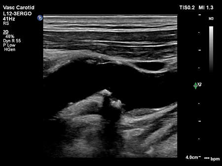
- Affiniti VM10.0 eL18-4 Vasc Carotid Verts CF
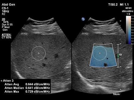
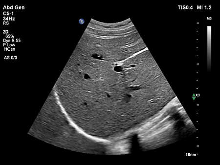
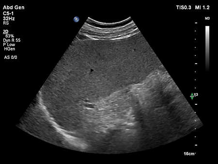
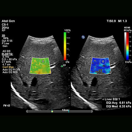
- Affiniti VM10.0 C5-1 Abd Gen Liver Fatty LFQ
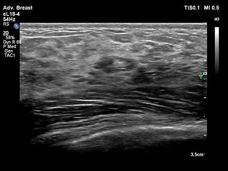
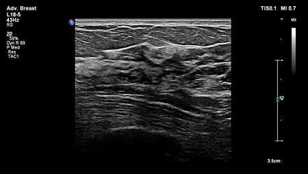
- Affiniti VM10.0 L12-5 Adv Breast 2D
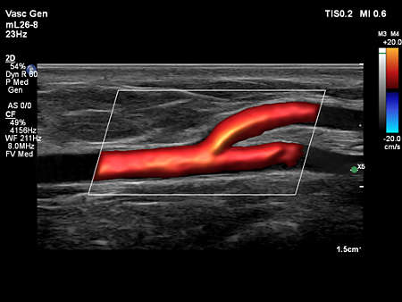
- Affiniti VM10.0 eL18-4 Vasc Carotid CCA CF
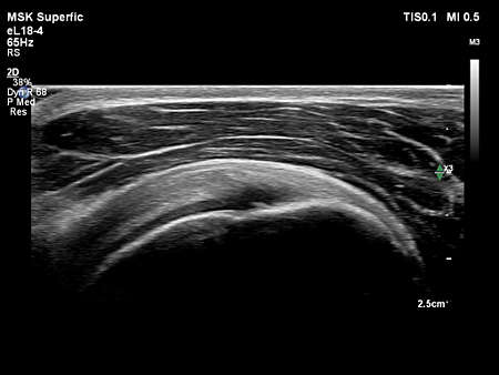
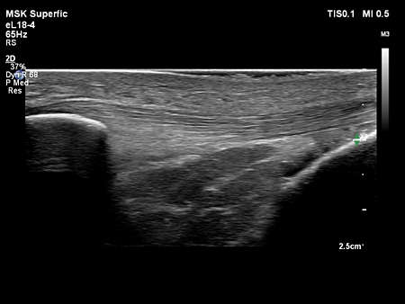
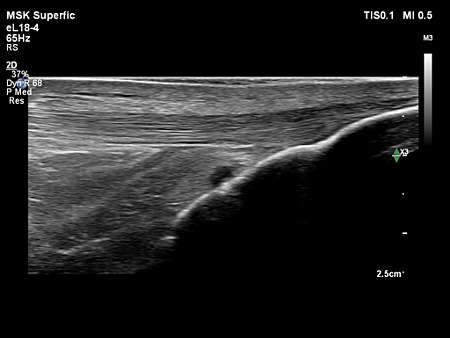
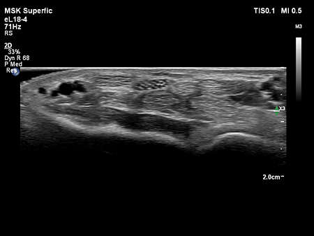
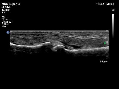
- Affiniti VM10.0 eL18-4 MSK Superfic Shoulder
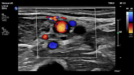
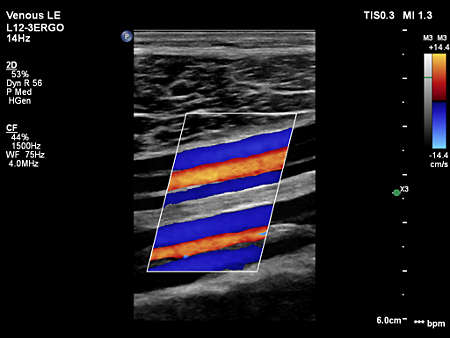
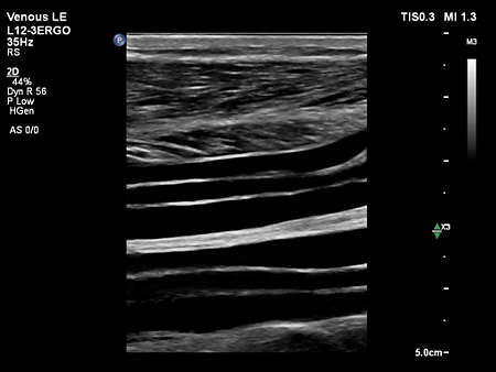
- Affiniti VM10.0 L12-3ERGO Venous LE Calf Veins 2D
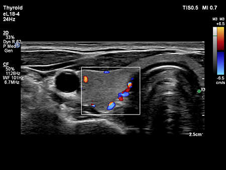
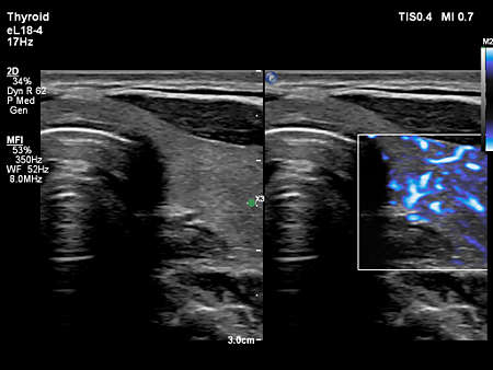
- Affiniti VM10.0 eL18-4 Thyroid CF
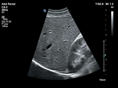
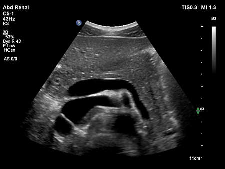
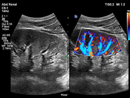
- Affiniti VM10.0 C5-1 Abd Renal Pancreas 2D
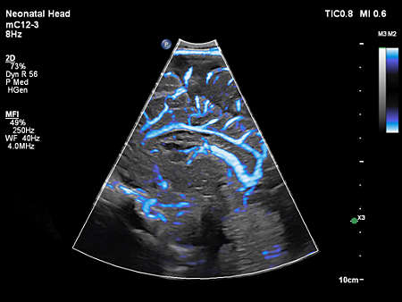
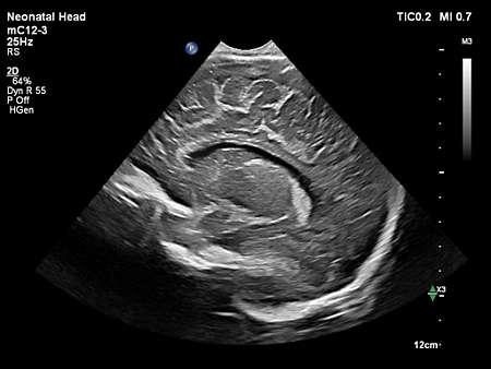
- Affiniti VM10.0 mC12-3 Neonatal Head CPA
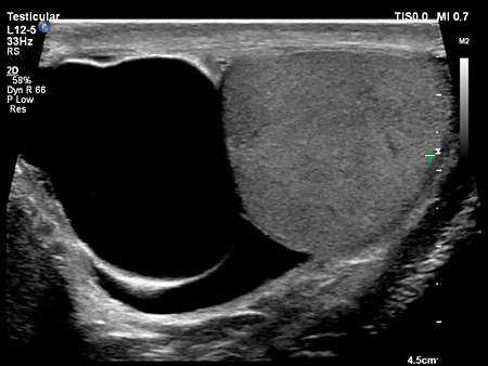
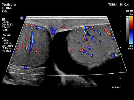
- Affiniti VM10.0 L12-5 Testicular Hydrocele
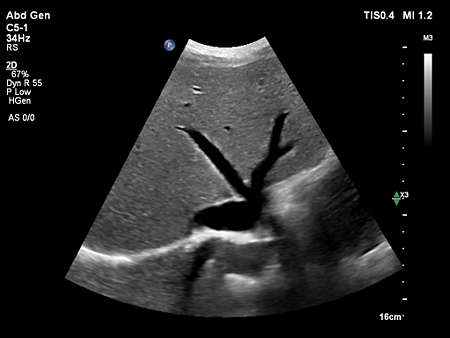
- Liver image from the Affiniti 70
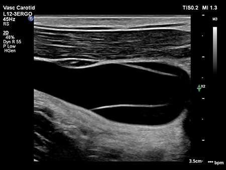
- Affiniti VM10.0 eL18-4 Vasc Carotid CCA 2D
- Affiniti Vm10.0 eL18-4 Thyroid MFI Compare
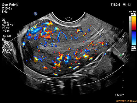
- Color Flow with Flow Viewer using C10-3v showing ovary perfusion
- Affiniti 30 ultrasound clinical image truevue
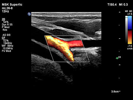
- mL26-8 paired with Flow Viewer, scanning on the General Vascular Preset
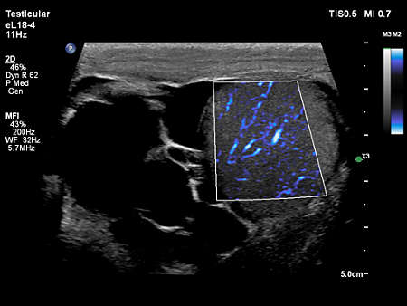
- Affiniti VM10.0 eL18-4 Testicular CF
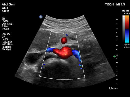
- Affiniti VM10.0 C5-1 Abd Gen IVC 2D
- Ovary MFI Flow Viewer
- mL26-8 paired with Flow Viewer, scanning on a calf vein
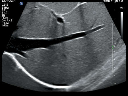
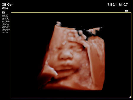
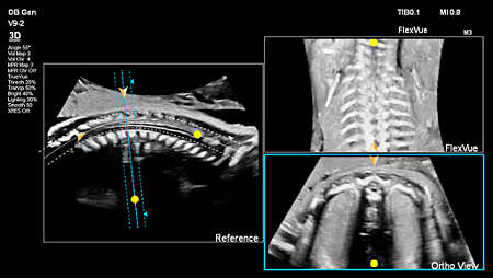
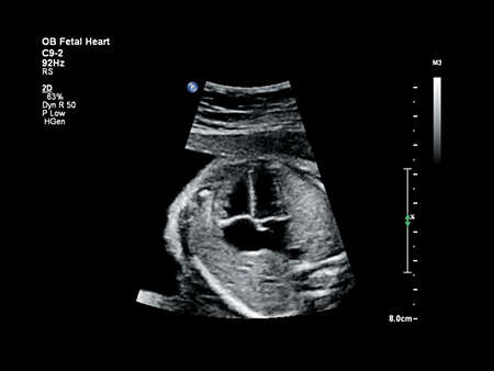
- Fetal profile shown using C9-2 transducer
- L12-3 paired with CEUS Auto Scan, scanning on the Vascular Carotid Preset.
- Affiniti VM10.0 C5-1 Abd Gen Liver 2D
- Affiniti VM10.0 C5-1 Abd Gen Liver Kidney 2D
- Aortic arch CPA with Flow Viewer using C9-2 transducer
- Affiniti VM10.0 L12-5 Adv Breast 2D
- Affiniti VM10.0 eL18-4 Thyroid 2D
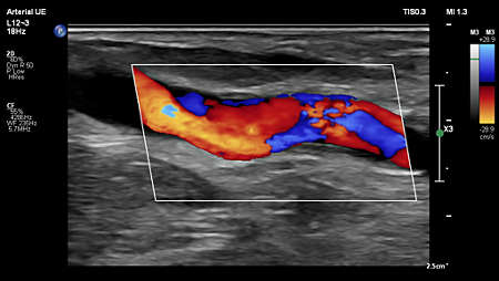
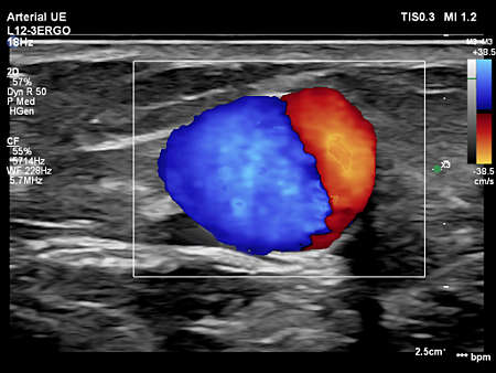
- Affiniti VM10.0 eL18-4 Arterial UE Fistual CF
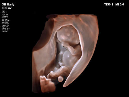
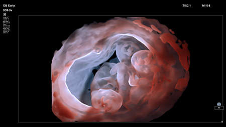
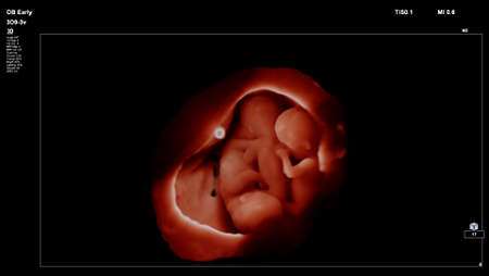
- Triplets shown using TrueVue on 3D9-3v transducer
- Affiniti VM10.0 eL18-4 MSK Superfic Nerve CF
- Affiniti 30 ultrasound clinical image truevue V6-2
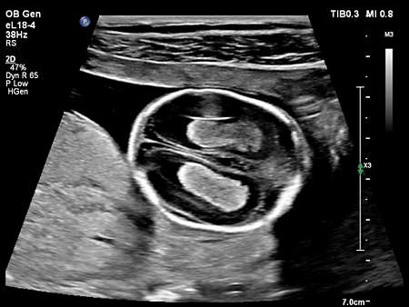
- Obstetric image from the Affiniti 70
- Affiniti VM10.0 C5-1 Abd Gen Liver Kidney HRI
- Affiniti VM10.0 C5-1 Abd Renal Kidney CPA Compare
- Affiniti VM10.0 L12-3 ERGO Venous LE Calf Veins CF
- Fetal Liver with MFI-eL18-4
- Auto Scan Of
- Affiniti VM10.0 L12-3 ERGO Vasc Carotid Verts 2D
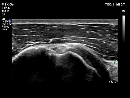
- Affiniti VM10.0 L12-5 MSK Gen Biceps Tendon
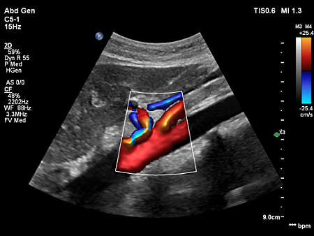
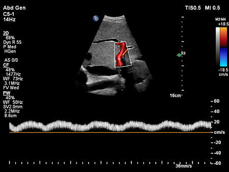
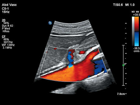
- Affiniti VM10.0 C5-1 Abd Gen Aorta FV
- Affiniti VM10.0 C5-1 Abd Gen Aorta Renals FV
- Triplets shown using GlassVue on 3D9-3v transducer
- Affiniti VM10.0 L12-3ERGO Arterial UE Fistual CF
- Uterus shown with MFI using C9-4v transducer
- Affiniti VM10.0 C5-1 Abd Gen FV Doppler
- Affiniti VM10.0 C5-1 Abd Gen Hepatic Veins 2D
- Affiniti VM10.0 eL18-4 Testicular Hydrocele
- Affiniti VM10.0 L12-5 Testicular Hydrocele
- Ovaries in MaxVue display using 3D9-3v transducer
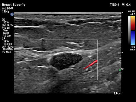
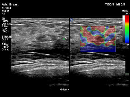
- eL18-4 scanning on a breast lesion.
- Gynecological image from the Affinti 70
- Affiniti VM10.0 eL18-4 Thyroid 2D
- Ovarian blood flow with MFI using C10-3v transducer
- Placenta CPA with Flow Viewer
- mL26-8 paired with Flow Viewer, scanning on a breast lesion
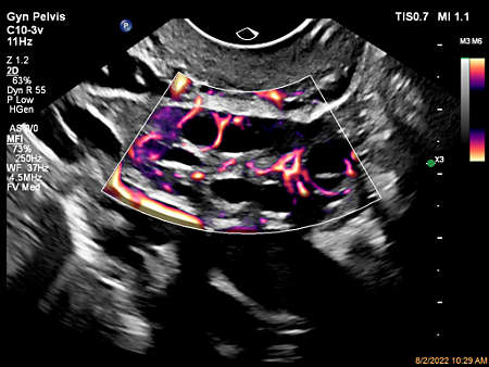
- Uterus Color Flow with Flow Viewer
- Affiniti VM10.0 L12-3ERGO Vasc Carotid Bulb Plaque
- Affiniti VM10.0 eL18-4 Arterial UE CF
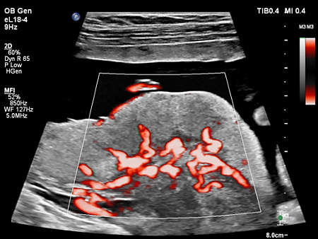
- Affiniti VM10.0 mC12-3 Neonatal Head MFI
- Flow Viewer applied to MFI with C10-3v uterus
- Affiniti VM10.0 eL18-4 Testicular Hydrocele
- L12-3 Ergo scanning on a calf vein
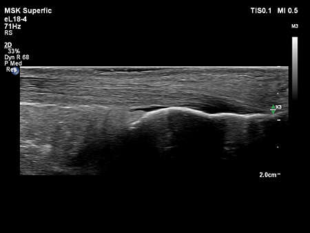
- Affiniti VM10.0 eL18-4 MSK Superfic Patellar Tendon
- Affiniti VM10.0 C5-1 Abd Gen Spleen 2D
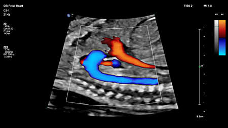
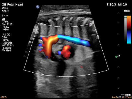
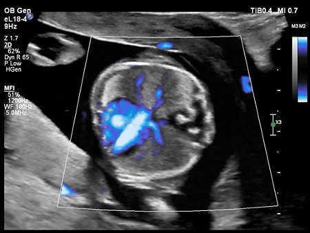
- Fetal Heart CPA with Flow Viewer
- mL26-8 paired with Flow Viewer, scanning on a thyroid
- Affiniti VM10.0 eL18-4 MSK Superfic Patellar Tendon
- Affiniti VM10.0 eL18-4 Adv Breast 2D
- Fetal Aortic Arch CPA with Flow Viewer
- Affiniti VM10.0 eL18-4 MSK Superfic Nerve
- Affiniti VM10.0 eL18-4 MSK Superfic Achilles Tendon
- Affiniti VM10.0 eL18-4 MSK Superfic Finger Pulley
- eL18-4 paired with FlowViewer, scanning on the Testicular Preset
- mL26-8 paired with Flow Viewer, scanning on a radial ulnar
- Affiniti VM10.0 L12-3ERGO Vasc Carotid IJV Valves
- Ovary Color Flow with Flow Viewer
- Fetal Lung Perfusion Color Flow with Flow Viewer
- Affiniti VM10.0 eL18-4 MSK Gen Shoulder 2D
- Affiniti VM10.0 eL18-4 Adv Breast Strain
- Affiniti VM10.0 eL18-4 Thyroid CF
- Affiniti VM10.0 mC12-3 Neonatal Head 2D
- Affiniti VM10.0 C5-1 Abd Gen Aorta Renal Vessels FV
- Gestational Sac in MaxVue display-C9-2
- Affiniti VM10.0 eL18-4 MSK Superfic Nerve Long
- Affiniti VM10.0 L12-5 Testicular Hydrocele
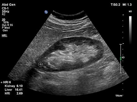
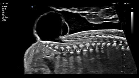
- Fetal abdomen image from the Affiniti 70
- Fetal heart image from the Affiniti 70


Features
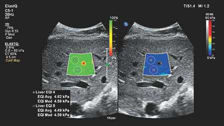
Auto ElastQ
Perform automated liver elastography with Auto ElastQ, and experience our next generation of liver health assessment. Auto ElastQ is designed to simplify user workflow with real-time, quantitative shear wave measurements.
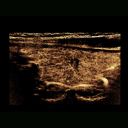
Contrast-enhanced ultrasound (CEUS)
CEUS can transform the role of ultrasound in the liver, allowing the study of the enhancement patterns of suspicious liver lesions in real time, as well as provide an alternative non-ionizing approach to the assessment of vesicoureteral reflux in pediatric patients.
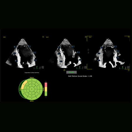
Auto Segmental Wall Motion Scoring
Provides automated evaluation of wall motion in a standard 17-segment bullseye display to aid objective LV wall assessment. With Auto SWMS, you can achieve greater reproducibility and efficiency in your workflows.
Specifications
- System dimensions
- Width
- 57.2 cm
- Height
- 142.2-162.6 cm
- Depth
- 98.3 cm
- Weight
- 83.6 kg
- Control panel
- Monitor size
- 54.6 cm
- Degrees of movement
- 180 degrees
Related products
- Choosing a new ultrasound system is all about balance. You need accurate diagnostic information quickly, a simplified yet intuitive user interface, and easy access to critical features, along with an ergonomic design and the latest technology.
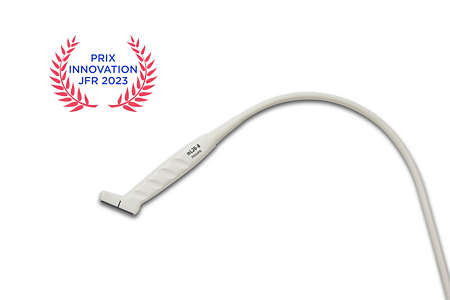
- Discover the award-winning Philips mL26-8 ultra-high frequency compact linear array transducer, designed to provide exceptional imaging versatility from head to hip. With specialized presets for MSK, breast, vascular, dermal, and ocular applications, the mL26-8 offers unmatched adaptability on EPIQ & Affiniti. Proud recipient of the 'Best Innovation Award in General Imaging' at Journées Francophones de Radiologie 2023.
Disclaimer
Available in select countries. Please consult your Philips representative for further details.
*based on a sample size of 20 users